Extension Agents get used to hearing that the local Extension Office is the community’s best kept secret. As much as we try to let folks know we’re here, many are still unaware of the services we provide. Even amongst the residents that are familiar with us, some of the services available remain unknown, especially our identification and diagnostic services. Here’s a rundown on some of the services available through your UF/IFAS Extension service.
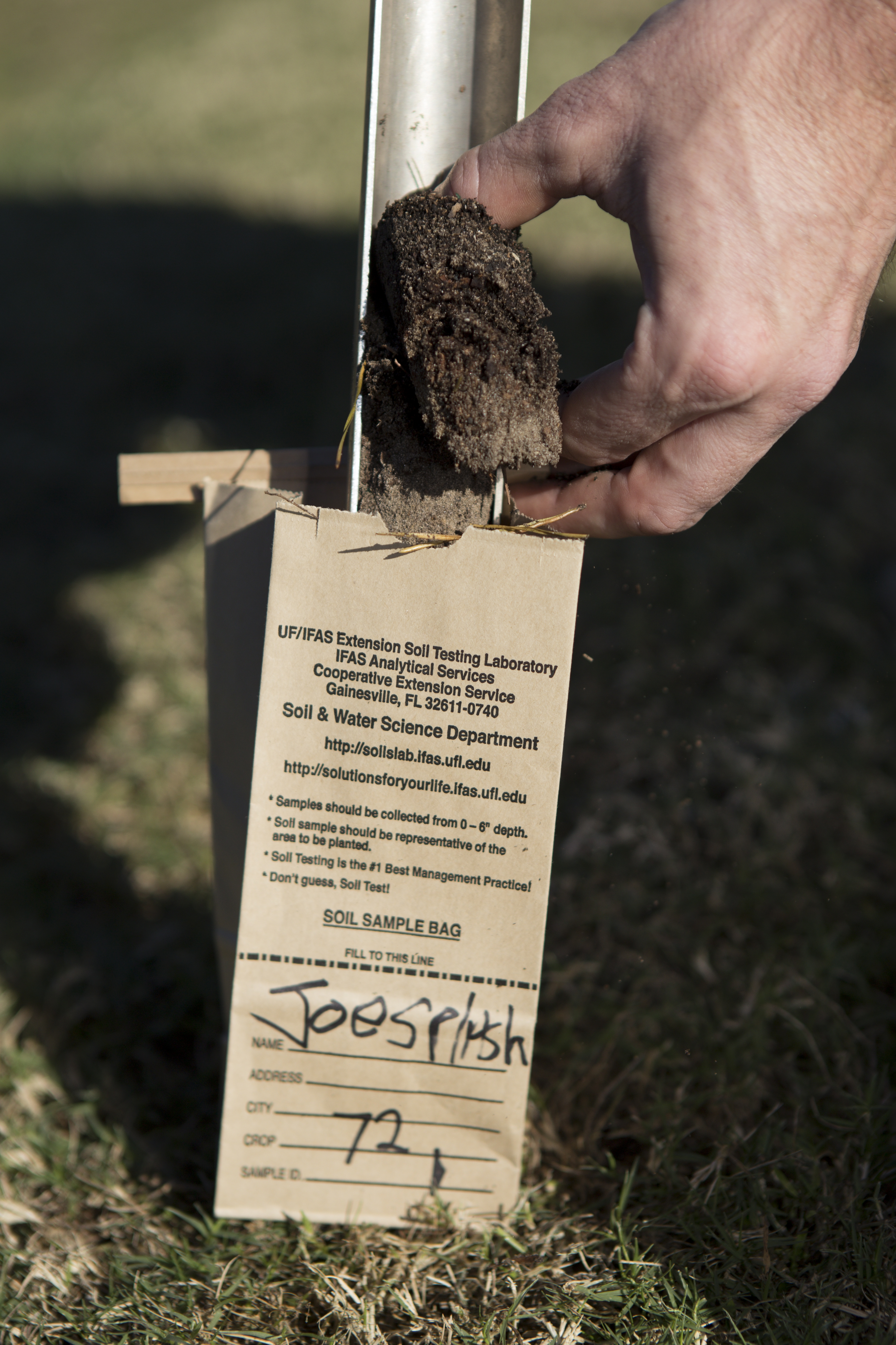
Taking a soil sample. UF/IFAS Photo by Tyler Jones
Soil Testing
This is probably our most well-known service, but it’s worth a reminder. For only $3 (pH only) or $10 (pH plus plant macro- and micro-nutrient values) per sample, plus shipping, you can have your soil analyzed in a state-of-the-art facility. To be clear, soil testing only provides a reading of your soil’s chemistry, specifically pH (acidity/alkalinity) and plant nutrient values. It does not provide information on any diseases or potential toxins that may be present in the soil. In addition to the results, you can specify the general type of plant you’re trying to grow (various grass species, vegetables, citrus, general trees and shrubs, etc.) and the report will provide recommendations to adjust the nutrient levels to be sure that plant is able to thrive. Your local agent receives a copy to help answer any questions you may have about the results or recommendations. More about soil and nutrient testing can be found at the Extension Analytical Services Laboratory website.
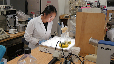
Experts at the Plant Disease Diagnostic Clinic can identify diseases present. Credit: UF/IFAS.
Plant Disease Diagnosis
UF/IFAS Extension has a great plant pathology lab on campus, but we also have a great resource close by in Gadsden County at the North Florida Research and Education Center’s (NFREC) Plant Disease Diagnostic Clinic. For a modest fee of $30, you can submit a sample of a diseased plant, and the lab manager will use the available methods to confirm the presence of disease and identify the disease-causing organism. Just like with the soil test results, you are provided with a recommendation on how to best treat the disease. The NFREC Plant Disease Diagnostic Clinic website has submittal forms, contact information, and directions for collecting a quality sample.
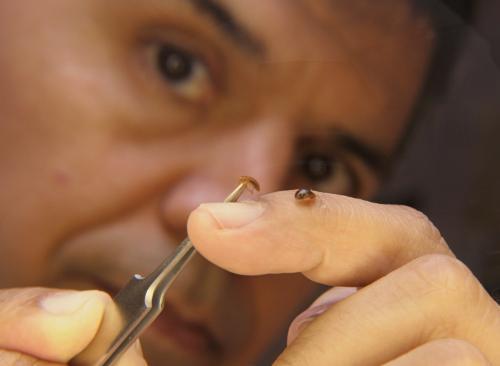
Need help with insect id? The DDIS system can help. Credit: UF/IFAS.
Plant and Insect Identification
While your local extension agent enjoys receiving plant and insect identification, there is an online submittal option available to use as well through our Distance Diagnostic Identification System (DDIS). You can set up an account and then upload photos of plants, insects, mushrooms, even diseased plants, and an expert on UF’s campus will do their best to identify it for you. The DDIS website has more information to help you set up a user account.
The Florida Cooperative Extension Service has many ways to help Florida citizens diagnose their landscape issues using science-based methods conducted by experts in state-of-the-art facilities. The above services are just a selection of the diagnostic capabilities available. To see a complete list, visit the IFAS Diagnostic Services website. You can always contact your local extension office, too, for assistance in identifying plants and insects, as well as diagnosing diseases.
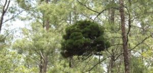
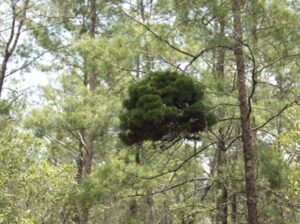
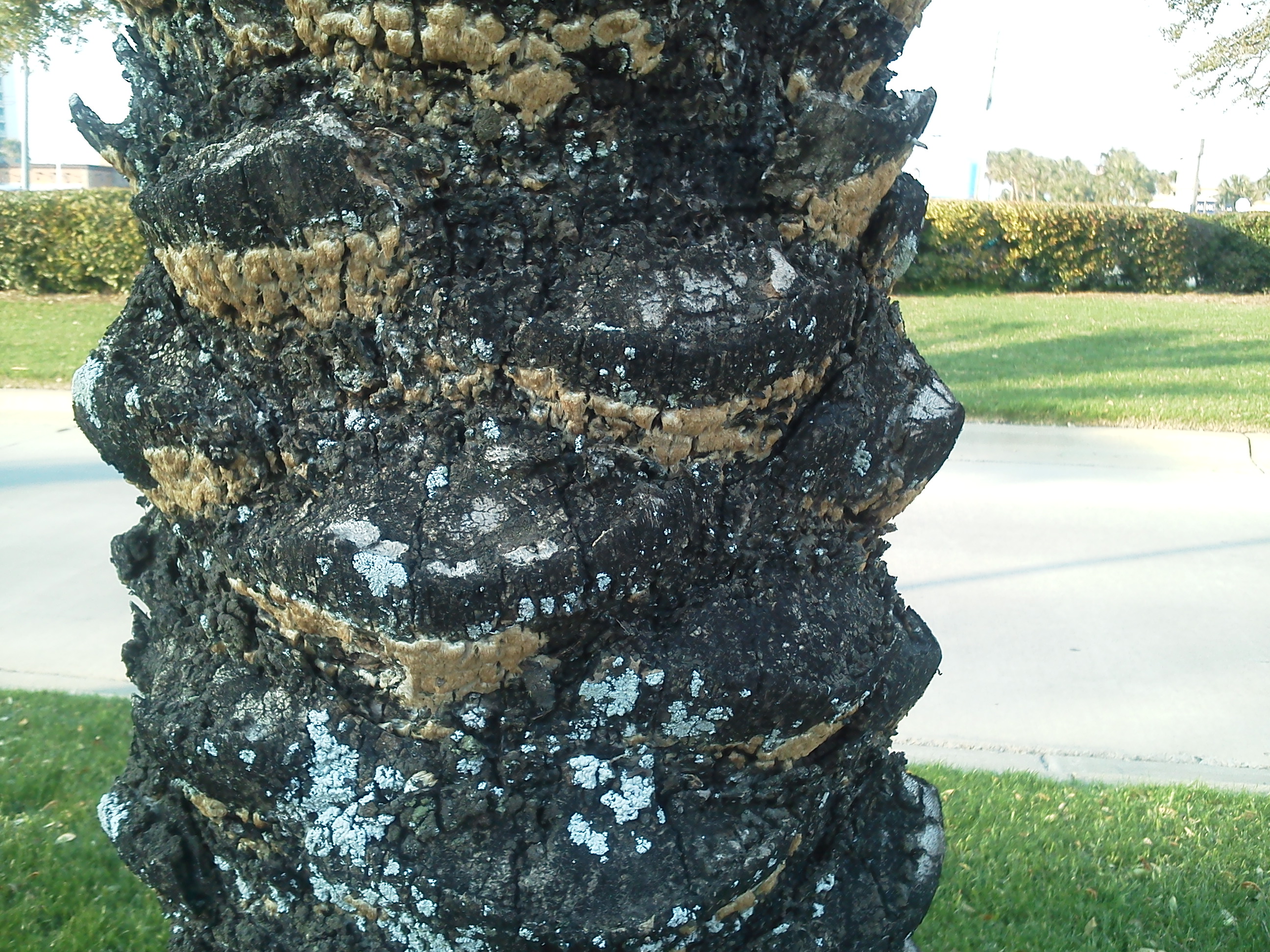
The telltale intense growth of a witches’ broom in a pine tree. Photo credit: Keith LeFevre
Our topic today might seem better suited to late October, but it can be observed in the woods year-round. During a recent Master Naturalist class, we discussed the various species of pines that grow in northwest Florida. All seven Florida native species—longleaf, loblolly, pond, slash, shortleaf, sand, and spruce—grow in our area of the state. While they can be differentiated based on growing location, needle length, and growth pattern, one of our class members had seen something really bizarre in the local pines.
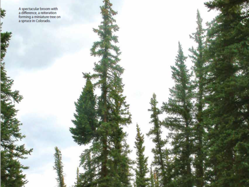
A witches’ broom in this spruce tree has resulted in a miniature version growing along its primary trunk. Photo credit: American Conifer Society
What he described was essentially an intense burst of pine needle growth at the tip of a branch. It stands out as deep green, dense, and unusual among the regular growth pattern of needles. The end result is essentially the production of a “mini-me,” a miniature copy of the normally growing tree, hanging off one of the branches. That afternoon while touring Blackwater River State Forest with a professional forester, we asked him about the strange phenomenon. He’d seen it many times and referred to it as a “witches’ broom.”
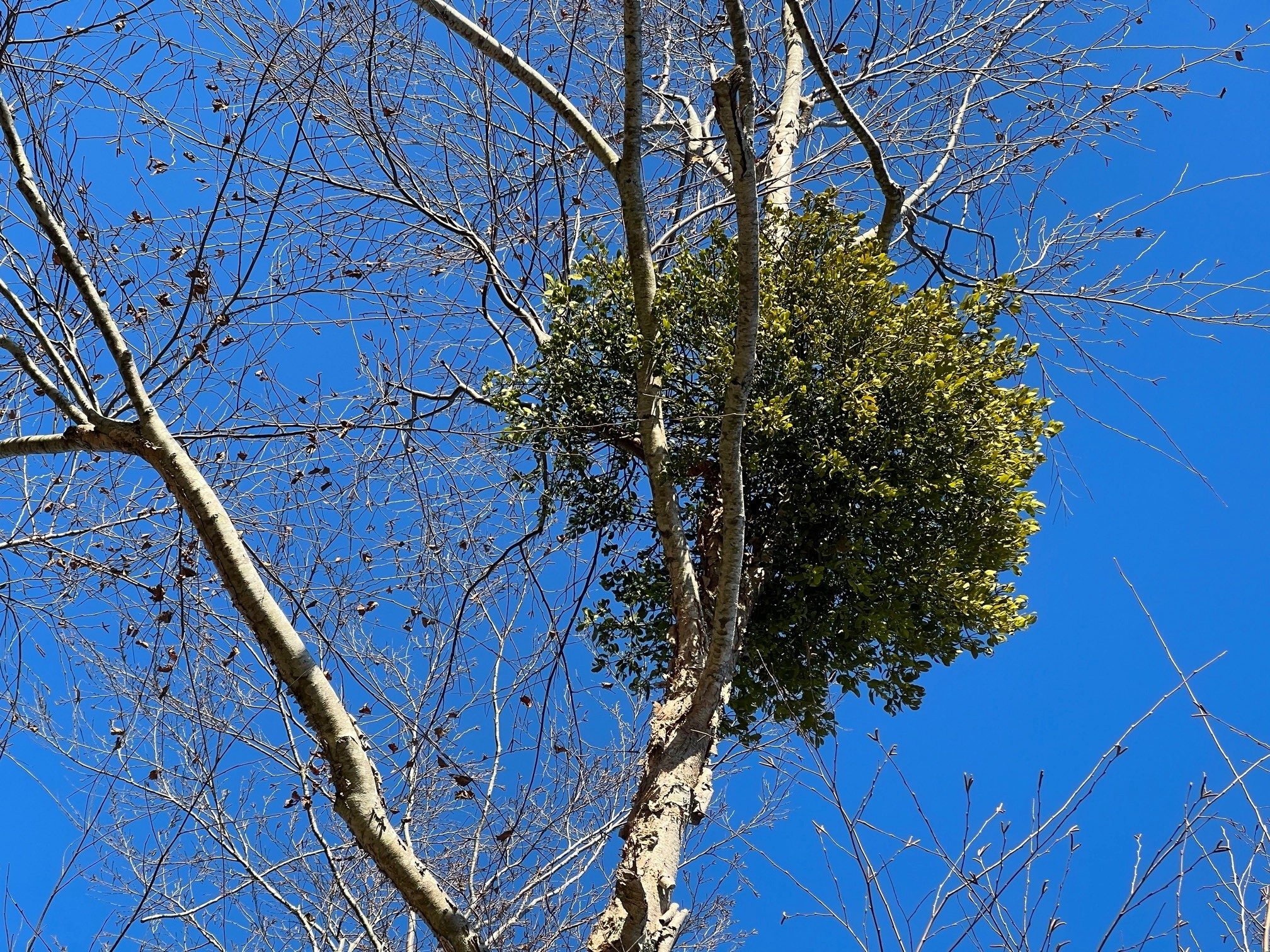
Mistletoe growing in a tree results from the same type of auxin disturbance as witches’ brooms. Photo credit: Carrie Stevenson, UF IFAS Extension
In normal tree growth, the trunk produces hormones called auxins, which control the division, expansion, and differentiation of cells. The hormones are concentrated in the growing tips of roots and shoots, and auxins maintain normal growth and keep smaller branches from overtaking the “leader.” Unusual growth occurs when the presence and concentration level of auxin is interfered with by an outside factor. The intense growth seen in these affected trees may be triggered in several ways, including pest, fungus, or mistletoe infestation, or death of terminal buds by environmental conditions. Phytoplasmas—bacteria that infect the phloem tissues—transferred by insect vectors (usually leafhoppers) are also blamed for the odd growth in some plants. Pines aren’t the only species affected; witches’ brooms can be found in other conifers like firs and junipers, nut species like hickory, pecan, and walnut, or in ashes, peaches, and elms.
The prolific growth of witches’ brooms is of great interest to horticulturists hoping to propagate dwarf varieties of the trees. This post by the American Conifer Society goes into great detail on how to “hunt”, cultivate, and encourage the growth of witches’ brooms into dwarf plants for the home landscape. Ecologically, witches’ brooms are not a huge problem for their host trees. Unless vulnerable to a massive outbreak of parasitic mistletoe, trees usually continue growing around them and live normal lifespans. The dense brush can even benefit wildlife, becoming a ready-made nest for birds or tree-dwelling mammals.
While palms may survive, or even thrive, for years in climates cooler than those to which they are native, eventually they may experience temperatures cold enough to cause injury. The January 21st snow and wind chill is likely to take a toll on many of the palms in Northwest Florida. We have experience with this since it also happened in January 2014 and December of 2022. Unfortunately, much of the damage in 2014 was not evident for 18-24 months and we are not out of the window for 2022. Healthy palms can hold on with stored food reserves, but repeated events can continue to weaken them. When cold damage is severe, plant tissues are destroyed and water uptake into the plant may be reduced for years. Many times, it is only the protected bud that will remain alive. These palms can still be saved if the bud remains alive. Winter is not over, even though the temperatures are now creeping higher and higher. So, evaluate what damage you may already have and prepare for any additional, yet to come. Here’s a reminder of what to do.
One of the most common problems associated with freezes is that the freeze-killed lower portion of the spear leaf is degraded by secondary fungi and bacteria that are always present in our natural environment. Palm owners are often anxious to trim off the damaged leaves following a cold weather event. Avoid the temptation to remove these fronds until danger of additional freezes has passed. Even dead leaves provide insulation to the critical bud. As the weather warms, the dead fronds need to be removed from around the bud so that the spear can begin to dry out. Drenching the bud area with a copper fungicide will reduce the secondary microbes. Repeat applications will need to continue as the palm leaves develop. Copper fungicides, unlike other fungicides, are active against bacteria and fungi. Be cautious to not use a copper nutrient spray rather than a fungicide. Delay fertilizer application until new fronds have developed. The best analysis for palms is 8-2-12 + 4Mg. Utilization of proper palm fertilization can improve cold hardiness of palms.
Palm trunk decay appeared 2 years after 2014 freeze.
Palms damaged by cold can still show symptoms six months to three years following a freeze. New leaves in the spring may appear misshapen. Usually, the palm will outgrow the damage. However, sometimes the palm loses its ability to take up water. If there is a sudden collapse of the fronds in the crown during the first hot days, the palm may die. There is nothing that can be done to save the palm.
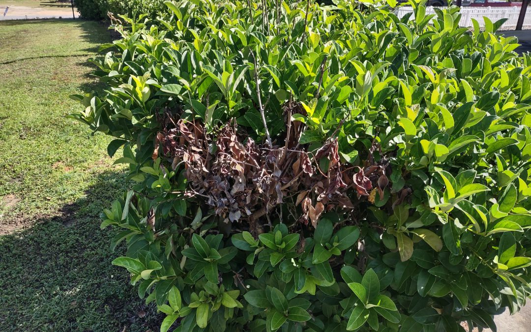
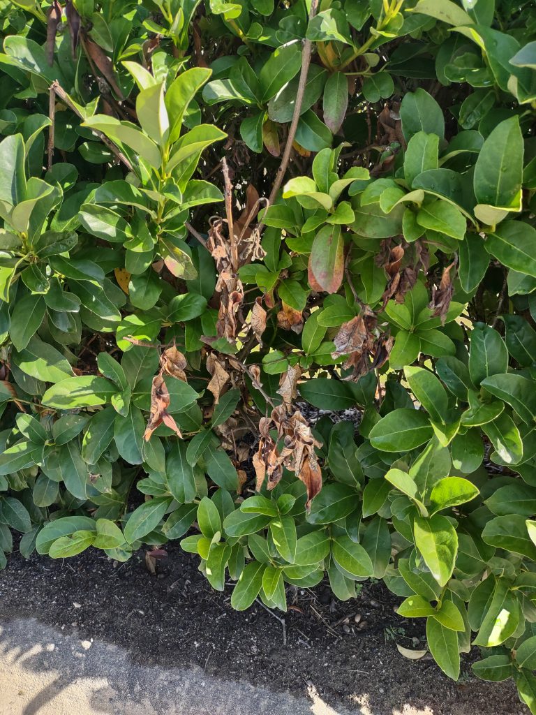
Sweet Viburnum (Viburnum odoratissimum) is thought of as being an ironclad landscape shrub, generally a rapid, healthy grower free of insects and disease. However, this spring, many Sweet Viburnum specimens across the Panhandle have experienced varying degrees of dieback, from individual shoots to entire sections of shrubs, caused by the fungal pathogen Botryosphaeria – commonly known as Bot Rot.
Typical symptom of Bot Rot on Sweet Viburnum. Photo courtesy of Daniel Leonard.
Bot Rot almost always appears after some kind of major stress event that impacts susceptible plants – drought, pruning wounds, nutritional deficiencies, or another environmental stress. We haven’t been afflicted lately with any serious drought conditions and the disease occurrences are too widespread to have been a result of isolated pruning or poor plant nutrition. However, the Panhandle did experience a major environmental event around Christmas 2022 that was plenty stressful for landscape plants, a weeklong Arctic blast of extreme cold. This abrupt hard freeze event in an otherwise mild winter is my best guess for what brought about the increased incidence of Botryosphaeria we have experienced this spring.
The Botryosphaeria fungus enters plants via wounds – in this case one probably caused by cold – and begins destroying the plant’s vascular system in the area. As the pathogen progresses, it eventually causes sunken cankers to appear, girdles the affected branch, and cuts off “circulation” in that stem. The first symptom of Bot Rot that gardeners notice is shoots rapidly wilting and exhibiting a blighted appearance, with brown, dead leaves holding onto affected limbs. Unfortunately, dieback isn’t always limited to individual shoots and can spread back into plants to eventually encompass whole branches. Entire plants dying from Bot Rot infection is not uncommon.
While there aren’t any fungicides that are effective in controlling or preventing Bot Rot, gardeners can arrest its spread by pruning out infected branches. To completely rid the plant of the fungus, make sure to prune 4” or so below the last infected plant tissue (symptomatic tissue will appear dark and discolored; healthy tissue will appear light and greenish). After pruning each affected plant, it is important to sanitize pruning equipment with either a 10% bleach solution or 70%+ isopropyl alcohol to prevent spreading pathogens to other healthy plants! Plants that have been irreparably disfigured by Bot Rot or outright killed may be pulled and discarded offsite.
While this year’s Bot Rot infestation has been extremely frustrating and similar future freeze events can’t be ruled out, gardeners should not give up on Sweet Viburnum, an excellent specimen or screening shrub. Keeping plants healthy with proper pruning, good fertility, and adequate irrigation is the best defense to ward off future infection when we experience harsh environmental conditions! For more information on Bot Rot, Sweet Viburnum, or any other horticultural topic, contact your local UF/IFAS County Extension Office! Happy Gardening.
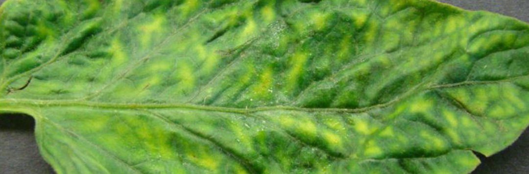
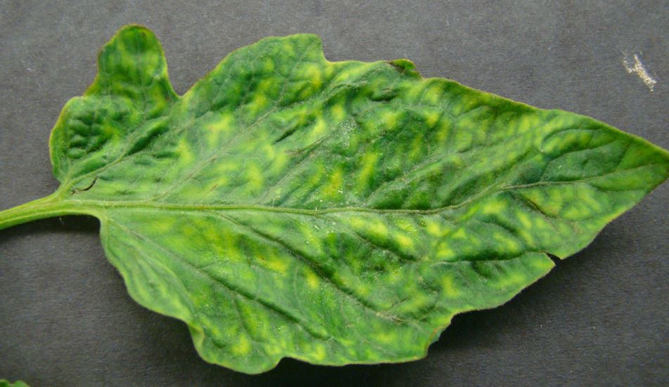
Did you know that the first virus discovered was in plants, not in humans? As early as 1857, tobacco farmers in the Netherlands recognized a new disease of tobacco. It wasn’t called a virus at the time as the causal agent was unknown. In 1886, Adolph Mayer, a German agricultural chemist, determined that the “tobacco mosaic” disease could be transmitted to healthy plants by rubbing them with infected leaf sap.
Tomato leaf with tobacco mosaic virus. Photo credit: UF Plant Pathology Department
When taking a plant pathology course in college, it amazed me that viral organisms were ever discovered. They are extremely small. So small that it was not until the development of the electron microscope in the late 1930s that scientists were able to see the structure of the tobacco mosaic virus. Viruses are 20 – 250 nanometers in diameter, about 100 times smaller than bacteria.
The discovery of fungi and bacteria came before the discovery of viruses as the cause for plant diseases. In most cases, we have many chemical options for control of fungal and bacterial diseases in plants. But there are few to no chemical options to control viral organisms in plants. By the nature of how a virus operates in a plant cell, chemical control results in death of the host cell, surrounding tissue and possibly the whole plant.
Control of viruses in plants involves eliminating the source of the virus such as nearby weeds, control of insect vectors that transmit the disease such as aphids and thrips and use of resistant plants.
Historically, plant diseases have caused major impacts on humans. In 1845, the Potato Famine in Ireland was caused by the fungus Phytophthora infestans. This disease was responsible for the death of more than 1 million people as it devastated the production of the potato as a major food source at the time and is credited for the beginning of plant pathology as a science. Cryphonectria parasitica, an introduced fungus, essentially wiped out the American chestnut as the dominant tree in the eastern U.S. forests during the early 1900s. The excellent wood was used to build homes and the nutritious nuts were eaten by humans and fed to livestock. Currently, UF/IFAS plant pathologists are working to solve Citrus Greening, a bacterial disease that has severely damaged the citrus industry and has the potential to completely eliminate citrus production in Florida.
If it were not for the land-grant university system, of which Extension Agents are a part, there would be few to no plant pathologists in the United States. It is these land-grant universities, like the University of Florida and others, that provide plant pathology courses, training, research, development and ultimately that graduate with degrees those who go on to careers in the field of plant pathology, discovering new diseases and developing controls for these diseases.